
A subgrain boundary is a wall of dislocations. The two ideal end members differ in where the misorientation axis points:
- a wall of edge dislocations rotates the lattice about an axis in the boundary plane - a tilt boundary
- a wall of screw dislocations rotates it about the boundary normal - a twist boundary
Real boundaries can contain both characters. This page computes the misorientation axes of the subgrain boundaries in a map and asks what they reveal. It ends with what a two-dimensional section can and cannot decide.
The page assumes that the map has already been divided into grains. Subgrain Boundaries explains the two reconstruction thresholds used below. Grain Boundary Properties introduces the per-segment properties, and Misorientation Theory develops the angle-axis description.
close all;

% load the map in its specimen plotting frame
plottingConvention.default('y↑→x');
mtexdata forsterite silent
Reconstruct and display the subgrain walls
The 1 degree threshold marks an inner boundary. The 15 degree threshold separates grains, while 'minPixel' removes indexed grains containing fewer than five measurements.
A raw boundary follows the square measurement grid, so its trace points mostly along the grid axes. Smoothing removes that staircase before the trace direction is interpreted. The flags 'noSimplify' and 'noRefine' retain every segment and its link to the two neighboring measurements through ebsdId.
% reconstruct low- and high-angle boundaries
[grains,ebsd] = calcGrains(ebsd,'threshold',[1*degree,15*degree],...
  'minPixel',5);

% smooth traces without breaking the segment-to-pixel relation
grains = smoothBoundary(grains,5,'noSimplify','noRefine');

% set up the IPF colouring
cKey = ipfColorKey(ebsd('fo').CS.properGroup);
cKey.ipfDirection = yvector;
color = cKey.orientation2color(ebsd('fo').orientations);

% draw the forsterite phase and both kinds of boundary
plot(ebsd('fo'),color,'faceAlpha',0.8,'figSize','large')
hold on
plot(grains.boundary,'lineWidth',2)

% fade the smallest boundary rotations and cap opacity at one
alpha = min(grains('fo').innerBoundary.misorientation.angle / ...
  (5*degree),1);
plot(grains('fo').innerBoundary,'lineWidth',1.5,...
  'edgeAlpha',alpha,'edgeColor','blue');
hold off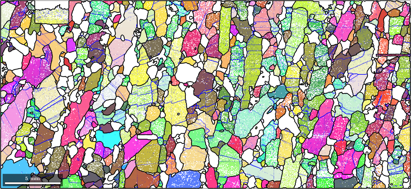
The blue lines are the subgrain boundaries. Rotations of 5 degrees and above are fully opaque, while smaller rotations fade towards the map. The lines are not scattered at random. They form families across the interiors of larger grains, which is the geometry expected of dislocation walls.
Misorientation axes in crystal coordinates
A reference frame is the coordinate system in which data are expressed. The crystal frame is fixed to the lattice basis. In this frame, the axis describes a lattice direction. For an ideal tilt wall it is the line direction of the edge dislocations that build the wall.
% extract the forsterite subgrain boundaries
subGB = grains('fo').innerBoundary;

% plot their axes in the fundamental sector
plot(subGB.misorientation.axis,'fundamentalRegion','figSize','small')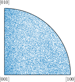
The scatter is too dense to show whether any directions are preferred. A density estimate of the same axes and its local maxima make the clustering visible.
% estimate and plot the crystal-frame axis density
crystalAxisDensity = calcDensity(subGB.misorientation.axis,...
  'halfwidth',3*degree);
plot(crystalAxisDensity,'figSize','small')
mtexColorbar('title','multiples of uniform density')

% report the two preferred crystal directions
[~,preferredCrystalAxes] = max(crystalAxisDensity,'numLocal',2);
preferredCrystalAxes = round(preferredCrystalAxes)
preferredCrystalAxes = Miller (Forsterite)
size: 2 × 1
antipodal: true
h k l
0 0 1
0 7 1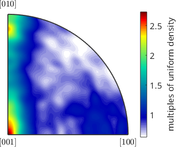
The two maxima are (001) and (071): rotations about the c axis and about an axis close to b. The clustering itself is the result. A few kinds of dislocation wall account for much of the subgrain-boundary population in this rock.
Attributing either axis to a slip system is a further step. It is a question about the material rather than the data alone. Naming the system requires knowing which dislocation systems can be active in olivine under the conditions experienced by this rock.
A limit of low-angle EBSD axes
The direction of a rotation axis becomes poorly constrained as its angle approaches zero. A small orientation error can then produce a large axis error. This page therefore uses clusters containing many segments and looks for continuity along walls. Do not interpret the axis of one faint, one-segment feature as evidence for a dislocation system.
Misorientation axes in specimen coordinates
The specimen frame is fixed to the sample and supplies the coordinates of this map. Expressed in that frame, the same axes show how the rotations associated with the walls are oriented in the specimen. This can relate to flow geometry, whereas the crystal-frame axes relate to the lattice.
A misorientation alone is a crystal-to-crystal rotation and contains no specimen-frame information. The orientations on both sides of each segment are needed, and ebsdId leads back to them.
oriGB = ebsd('id',subGB.ebsdId).orientationsoriGB = orientation (Forsterite → y↑→x)
size: 31803 × 2The displayed size has one row per segment and two columns for its two sides. axis computes their axes in the specimen frame.
axS = axis(oriGB(:,1),oriGB(:,2),'antipodal')

% plot the specimen-frame axes
plot(axS,'MarkerAlpha',0.2,'MarkerSize',2,'figSize','small')
axS = vector3d (y↑→x)
size: 31803 × 1
antipodal: true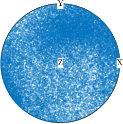
The flag 'antipodal' is essential because the sides of a boundary have no intrinsic order. Swapping them inverts the misorientation and reverses its axis, but the physical axis has not changed.
The scatter still overlaps heavily, so estimate its density as before.
% estimate and plot the specimen-frame axis density
specimenAxisDensity = calcDensity(axS,'halfwidth',5*degree);
plot(specimenAxisDensity,'figSize','small')
mtexColorbar('title','multiples of uniform density')

[~,preferredSpecimenAxis] = max(specimenAxisDensity)
annotate(preferredSpecimenAxis)
preferredSpecimenAxis = vector3d (y↑→x)
antipodal: true
x y z
0.195 0.706 0.68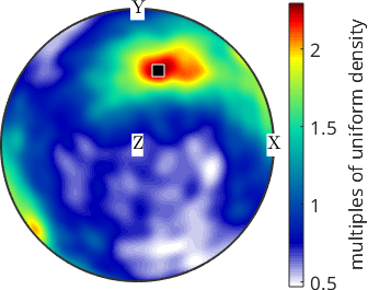
The maximum lies obliquely between TD and ND, with a smaller RD component. The specimen history is not supplied with this example, so the direction should not be assigned to a loading or flow mechanism here.
What a two-dimensional section can decide
A polished section does not reveal the boundary plane. It reveals only the trace, the line where that plane intersects the section. The boundary inclination remains unknown, so tilt and twist cannot generally be distinguished from a two-dimensional map. One part of the question can still be answered.
A twist boundary has its misorientation axis along the boundary normal. That axis is perpendicular to every direction in the plane, including the trace. An axis parallel to the trace therefore rules out twist and makes tilt likely. An axis perpendicular to the trace leaves tilt, twist, and mixed character open because the trace is only one direction in the plane.
The map below colours every subgrain segment by the angle between its trace and specimen-frame axis. Blue means close to 0 degrees and therefore likely tilt. Red means close to 90 degrees and remains undecided. Colour saturation follows the misorientation angle, so that the least reliable axes fade towards neutral grey.
% compute the angle between each trace and its unoriented axis
traceAxisAngle = angle(subGB.direction,axS)./degree;

% desaturate the smallest rotations without changing their angle hue
traceAxisColor = num2rgb(traceAxisAngle,blue2redColorMap,...
  'range',[0 90]);
traceAxisColor = alpha .* traceAxisColor + (1-alpha) .* 0.65;

plot(ebsd('fo'),color,'faceAlpha',0.5,'figSize','large')
hold on
plot(grains.boundary,'lineWidth',2)
plot(subGB,traceAxisColor,'lineWidth',2)
hold off
mtexColorMap blue2red
setColorRange([0 90])
mtexColorbar('title','axis-trace angle (degrees)')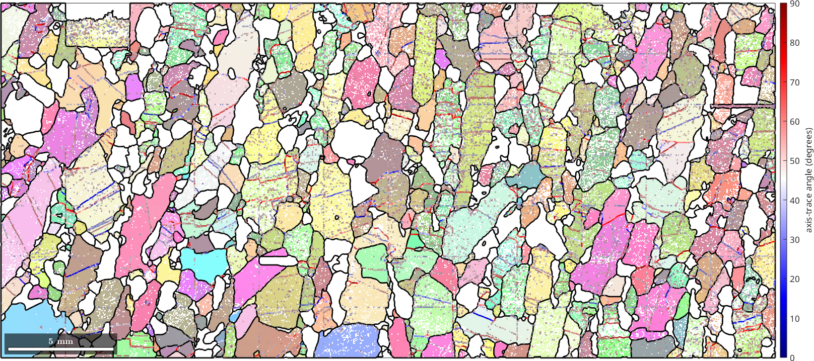
Both colours are present. A long, straight subgrain boundary generally holds one colour along its length, so its character can be read with more confidence. The intervening speckle is different: isolated one- and two-segment features can change colour from one segment to the next.
The following calculation makes that difference explicit. It computes the standard deviation within every connected component containing at least 20 segments, then averages those deviations with segment-count weights. The comparison value is the standard deviation over the whole map.
isWall = subGB.componentSize >= 20;
[~,~,wallId] = unique(subGB.componentId(isWall));
wallSize = accumarray(wallId,1);
componentScatter = accumarray(wallId,traceAxisAngle(isWall),[],@std);
withinWallScatter = sum(wallSize .* componentScatter) / sum(wallSize);
mapScatter = std(traceAxisAngle);

fprintf('Mean within-wall scatter: %.1f degrees\n',withinWallScatter);
fprintf('Whole-map scatter: %.1f degrees\n',mapScatter);
Mean within-wall scatter: 19.4 degrees
Whole-map scatter: 22.5 degreesThe mean within-wall scatter is 19.4 degrees, against 22.5 degrees over the map. The difference is modest. Continuity along a long boundary is therefore more informative than the map-wide average.
Deciding the red boundaries individually requires their boundary planes, which means three-dimensional data. A statistical argument over many traces can instead recover a distribution of planes, but not the plane of one segment. See Grain Boundary Normal Distribution for that population-level analysis.
References
- W. T. Read and W. Shockley, Dislocation Models of Crystal Grain Boundaries, Physical Review 78 (1950), 275--289. This is the classic dislocation model for low-angle boundaries.
- A. P. Sutton and R. W. Balluffi, Interfaces in Crystalline Materials, Oxford University Press, 2006. Chapter 1 develops the five macroscopic boundary parameters and the tilt and twist end members.
- G. E. Lloyd, A. B. Farmer and D. Mainprice, Misorientation Analysis and the Formation and Orientation of Subgrain and Grain Boundaries, Tectonophysics 279 (1997), 55--78. The paper combines misorientation axes with boundary traces in geological microstructures.
- D. J. Prior, Problems in Determining the Misorientation Axes, for Small Angular Misorientations, Using Electron Backscatter Diffraction in the SEM, Journal of Microscopy 195 (1999), 217--225. The paper quantifies the loss of axis precision at small angles.
Next
CSL Boundaries is the next page in this chapter and classifies special high-angle relationships. For the statistical recovery of boundary planes from traces, continue with Grain Boundary Normal Distribution.
Citing this page.
This page is part of the documentation of
MTEX, a free and open
source MATLAB toolbox for analyzing and modeling crystallographic textures.
It was written by The MTEX Developers and is published at
https://mtex-toolbox.github.io/TiltAndTwistBoundaries.html.
If you use MTEX, or reuse text or figures from this page, in your research,
please cite
F. Bachmann, R. Hielscher, H. Schaeben: Texture Analysis with MTEX - Free and Open Source Software Toolbox, Solid State Phenomena 160 (2010), 63-68. 10.4028/www.scientific.net/SSP.160.63
BibTeX
@article{bachmann2010mtex,
  author  = {F. Bachmann and R. Hielscher and H. Schaeben},
  title   = {Texture Analysis with MTEX - Free and Open Source Software Toolbox},
  journal = {Solid State Phenomena},
  volume  = {160},
  pages   = {63-68},
  year    = {2010},
  doi     = {10.4028/www.scientific.net/SSP.160.63},
  url     = {https://doi.org/10.4028/www.scientific.net/SSP.160.63}
}
Other papers describing specific MTEX methods are listed under Publications — please cite the one that best fits your application. The MTEX source code is licensed under the GNU General Public License v2.0; the text and figures of this documentation are licensed under CC BY 4.0, which permits reuse — including by automated systems — provided The MTEX Developers and this page are credited.