
A pole density function is a two-dimensional projection of an ODF. Fix a crystal direction \(\vec h\) and ask how much material points it along each specimen direction \(\vec r\).
This page assumes the ODF and mrd normalization introduced in ODF Theory. Crystal directions and their Miller indices are introduced in Miller Indices.
Formally, the pole density function is the ODF integrated over every orientation that maps \(\vec h\) onto \(\vec r\),
\[ P_{\vec h}(\vec r) = \int_{g \vec h = \vec r} f(g)\, \mathrm{d}g. \]
This integral is the spherical Radon transform of the ODF. It collapses a whole orientation fibre to one value, so a pole figure is a projection rather than a section through orientation space. One pole figure cannot determine a three-dimensional ODF, and different ODFs can produce the same pole figure.
Like the ODF itself, the result is a density in multiples of a random distribution (mrd). A value of 2 mrd means twice the random density at that specimen direction, not two percent of the material.
After background, defocusing, and structure-factor corrections, this density is proportional to what a diffraction experiment measures. Pole figures therefore connect diffraction measurements to texture; see Reconstructing an ODF for the inverse problem.
A plotting convention states how a reference frame is laid out on screen. The following convention draws specimen Y upward and specimen X to the right. It does not rotate the specimen or change the ODF.
plottingConvention.default('y↑→x');A Model Texture to Look At
The example combines two single-orientation components with one fibre component. Trigonal symmetry makes the difference between a direction and its opposite visible in the third pole figure.
cs = crystalSymmetry('32');
mod1 = orientation.byEuler(90*degree,40*degree,110*degree,'ZYZ',cs);
mod2 = orientation.byEuler(50*degree,30*degree,-30*degree,'ZYZ',cs);

odf = 0.2*unimodalODF(mod1) ...
  + 0.3*unimodalODF(mod2) ...
  + 0.5*fibreODF(Miller(0,0,1,cs),vector3d.X,...
  'halfwidth',10*degree);

% select three crystal directions in four-index notation
h = Miller({1,0,-1,0},{0,0,0,1},{1,1,-2,1},cs);
plotPDF fixes each crystal direction in h and draws its pole density over specimen directions. The panels use the default spherical projection.
plotPDF(odf,h);
mtexColorMap LaboTeX;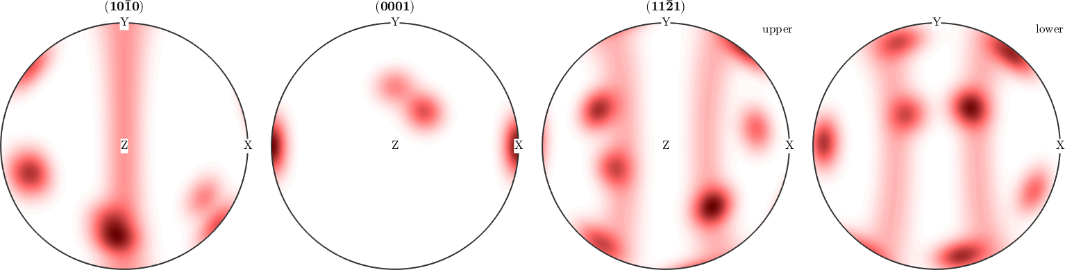
The two single-orientation components make localized spots. The fibre component makes a ring because its remaining rotational freedom becomes a curve in the pole figure.
MTEX draws only the upper hemisphere when the lower hemisphere contains no independent information. That is why the first two pole figures use one disc while the third uses two.
When the Two Hemispheres Differ
The 'complete' flag displays both hemispheres for every direction. It changes the displayed region; it does not symmetrize the pole density or restore information lost by the projection.
plotPDF(odf,h,'complete');
mtexColorMap LaboTeX;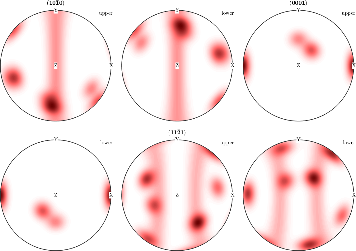
The upper and lower hemispheres agree in exactly three situations:
- the crystal direction \(\vec h\) is symmetrically equivalent to \(-\vec h\) - true here for \((10\bar10)\) and the c-axis \((0001)\), but not for \((11\bar21)\);
- the crystal symmetry contains inversion and is therefore a Laue group;
- antipodal symmetry is assumed, as is conventional for kinematic diffraction under Friedel's law; see Axes and Antipodal Symmetry.
Dynamical or resonant diffraction may distinguish the two sides. The antipodal assumption therefore belongs to the experiment, not to the definition of an ODF.
The 'antipodal' flag averages opposite directions. With 'complete' retained below, both equal halves remain visible for comparison.
plotPDF(odf,h(3),'antipodal','complete');
mtexColorMap LaboTeX;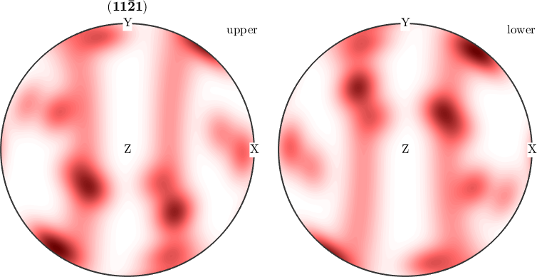
Opposite points in the two discs now have the same colour. Drawing both discs does not make them independent measurements.
Values Rather Than Pictures
calcPDF with only a crystal direction returns a spherical function. Passing a specimen direction as well evaluates that function at one point.
pdf = calcPDF(odf,Miller(1,0,0,cs))
densityAtX = calcPDF(odf,Miller(1,0,0,cs),vector3d.X)
pdf = S2FunHarmonicSym (y↑→x)
bandwidth: 25
antipodal: true
densityAtX =
0.1982The printed summary identifies pdf as a spherical function. Its value along specimen X is 0.1982 mrd, so this texture avoids that direction.
Averaged over the whole sphere, every normalized pole density function is 1 mrd, just as every normalized ODF has mean 1 mrd.
meanDensity = mean(pdf)meanDensity =
1.0000Following a Pole-Figure Maximum into Orientation Space
A pole-figure maximum still represents a whole orientation fibre. Find the strongest specimen direction for the third pole figure, then inspect the ODF along the corresponding fibre with plotFibre.
pdfPeak = calcPDF(odf,h(3));
[~,rPeak] = max(pdfPeak);
f = fibre(h(3),rPeak);

close all;
plotFibre(odf,f,'LineWidth',2,'figSize','small');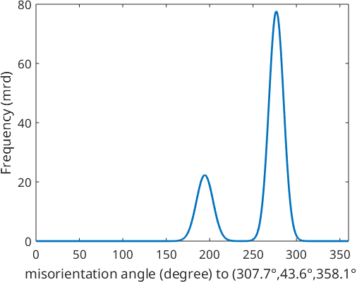
The line is the ODF density itself, not another projection. Its peaks locate the components that this fibre passes through, while their average along the curve is the pole-figure value at rPeak. This is the degree of freedom that the pole figure hides.
Further Reading
- H.-J. Bunge, Texture Analysis in Materials Science: Mathematical Methods, Butterworths, English ed., 1982. Chapters on pole figures and orientation distributions develop the classical forward and inverse transforms.
- ASTM International, ASTM E81-96(2024): Standard Test Method for Preparing Quantitative Pole Figures. It covers quantitative X-ray pole figures and the distinction between complete, partial, and calculated pole figures.
- H.-J. Bunge and C. Esling, Determination of the odd part of the texture function by anomalous scattering, Journal of Applied Crystallography 14 (1981), 253--255. It explains why ordinary diffraction loses the odd ODF part and how anomalous scattering changes that limitation.
Next
The other projection fixes a specimen direction instead of a crystal direction; continue with Inverse Pole Figures. Going the other way, from measured pole figures to an ODF, is Reconstructing an ODF.
Citing this page.
This page is part of the documentation of
MTEX, a free and open
source MATLAB toolbox for analyzing and modeling crystallographic textures.
It was written by The MTEX Developers and is published at
https://mtex-toolbox.github.io/ODFPoleFigure.html.
If you use MTEX, or reuse text or figures from this page, in your research,
please cite
F. Bachmann, R. Hielscher, H. Schaeben: Texture Analysis with MTEX - Free and Open Source Software Toolbox, Solid State Phenomena 160 (2010), 63-68. 10.4028/www.scientific.net/SSP.160.63
BibTeX
@article{bachmann2010mtex,
  author  = {F. Bachmann and R. Hielscher and H. Schaeben},
  title   = {Texture Analysis with MTEX - Free and Open Source Software Toolbox},
  journal = {Solid State Phenomena},
  volume  = {160},
  pages   = {63-68},
  year    = {2010},
  doi     = {10.4028/www.scientific.net/SSP.160.63},
  url     = {https://doi.org/10.4028/www.scientific.net/SSP.160.63}
}
Other papers describing specific MTEX methods are listed under Publications — please cite the one that best fits your application. The MTEX source code is licensed under the GNU General Public License v2.0; the text and figures of this documentation are licensed under CC BY 4.0, which permits reuse — including by automated systems — provided The MTEX Developers and this page are credited.