
X-ray, synchrotron and neutron diffraction measure many crystals at once. For each selected lattice plane, a pole figure records intensity over specimen directions. Each value combines all crystal orientations that place that plane normal in the measured direction.
This tutorial imports four pole figures, corrects their intensities, reconstructs an orientation distribution function (ODF), and checks the reconstruction against the measurements. New MTEX users may first want General Concepts. Miller indices are introduced in Miller Indices, and specimen symmetry in Specimen Symmetry.
Describe the experiment
Import pole figure data starts the import wizard. The wizard asks for scientific inputs that a numeric file may not contain, then writes a reproducible script. Import Pole Figure Data explains those choices in detail.
The point group of this ZnCuTi phase is 6/mmm. The lattice parameters and alignment options state how its lattice is expressed in the crystal frame. That alignment belongs to the frame, not to the symmetry itself.
CS = crystalSymmetry('6/mmm',[2.633 2.633 4.8],...
  'X||a*','Y||b','Z||c');
Specimen symmetry describes a physical invariance of the specimen. Choosing 1 imposes no such invariance on this reconstruction.
SS = specimenSymmetry('1');

% plotting convention: z out of the screen, x pointing north
plottingConvention.default('y←↑x');
The first four files contain the specimen measurements. The second four contain measurements of a texture-free reference specimen made with the same instrument.
pname = fullfile(mtexDataPath,'PoleFigure','ZnCuTi');

fname = {...
  fullfile(pname,'ZnCuTi_Wal_50_5x5_PF_002_R.UXD'),...
  fullfile(pname,'ZnCuTi_Wal_50_5x5_PF_100_R.UXD'),...
  fullfile(pname,'ZnCuTi_Wal_50_5x5_PF_101_R.UXD'),...
  fullfile(pname,'ZnCuTi_Wal_50_5x5_PF_102_R.UXD')};

fnameDef = {...
  fullfile(pname,'ZnCuTi_defocusing_PF_002_R.UXD'),...
  fullfile(pname,'ZnCuTi_defocusing_PF_100_R.UXD'),...
  fullfile(pname,'ZnCuTi_defocusing_PF_101_R.UXD'),...
  fullfile(pname,'ZnCuTi_defocusing_PF_102_R.UXD')};
Match files to lattice planes
Each Miller index must describe the reflection in the file at the same position. A wrong assignment still produces an ODF, but it gives the reconstruction the wrong physical measurement model.
h = {...
  Miller(0,0,2,CS),...
  Miller(1,0,0,CS),...
  Miller(1,0,1,CS),...
  Miller(1,0,2,CS)};
Import and correct the intensities
PoleFigure.load combines the four files in one PoleFigure object. Its display reports four pole figures with 1152 specimen directions each, for 4608 measured intensities in total.
pf = PoleFigure.load(fname,h,CS,SS,'interface','uxd')pf = PoleFigure (y←↑x)
crystal symmetry : 6/mmm, X||a*, Y||b, Z||c
h = (0002), r = 1152 × 1 points
h = (101̅0), r = 1152 × 1 points
h = (101̅1), r = 1152 × 1 points
h = (101̅2), r = 1152 × 1 pointsKeep the reference measurement quiet because it is only an input to the correction. correct divides the specimen data by this reference to compensate for intensity lost as the specimen tilts.
pfDef = PoleFigure.load(fnameDef,h,CS,SS,'interface','uxd');
pf = correct(pf,'def',pfDef);
Check the corrected data
Plot measurements before attempting an inversion. Check that each panel has the intended Miller index and that the plotted specimen axes match the experimental alignment. A plotting convention controls only where directions appear on screen; it does not repair a wrong reference frame.
plot(pf);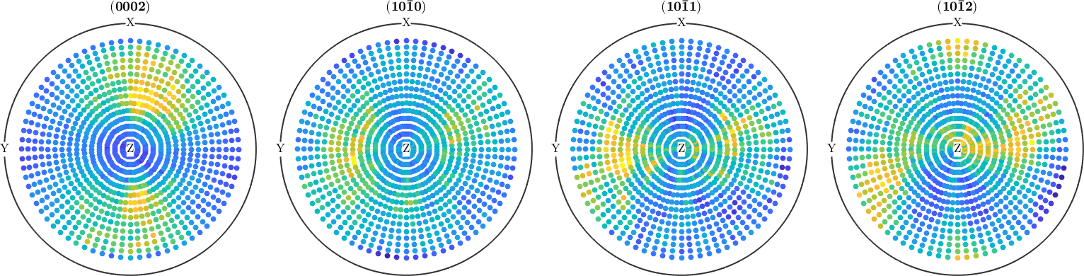
The four panels share the same sampling grid, while their broad intensity maxima occur at different specimen directions. Those distinct patterns provide independent constraints on the ODF.
Corrections can create negative intensities, which are not physical diffraction measurements. Count them before clipping them to zero.
numNegative = nnz(pf.intensities < 0)
pf(pf.intensities < 0) = 0;
numNegative =
0
numNegative is 0 for this dataset, so clipping changes no values and a second plot would be identical. Outlier removal, rotation, scaling, and other corrections are covered in Modify Pole Figures.
Reconstruct an ODF
calcODF finds an ODF whose recalculated pole figures fit the corrected measurements. 'silent' suppresses the solver iteration history, while the returned object remains visible.
odf = calcODF(pf,'silent')odf = SO3FunRBF (6/mmm → y←↑x)
uniform component
weight: 0.54
multimodal components
kernel: de la Vallee Poussin, halfwidth 5°
center: 9924 orientations, resolution: 5°
weight: 0.46The display identifies the result as an SO3FunRBF. Radial basis functions are its numerical representation, not a different scientific quantity. MTEX plotting and analysis commands operate through the common ODF interface.
Reconstruction is not unique. Distinct ODFs can have identical pole figures, even with perfect measurements. More independent pole figures constrain the result, but they do not remove the fundamental ambiguity. The Ghost Effect explains what the measurement cannot determine.
Check the reconstruction
Recalculate the four measured pole figures from the ODF and compare them with the corrected data above.
plotPDF(odf,pf.h);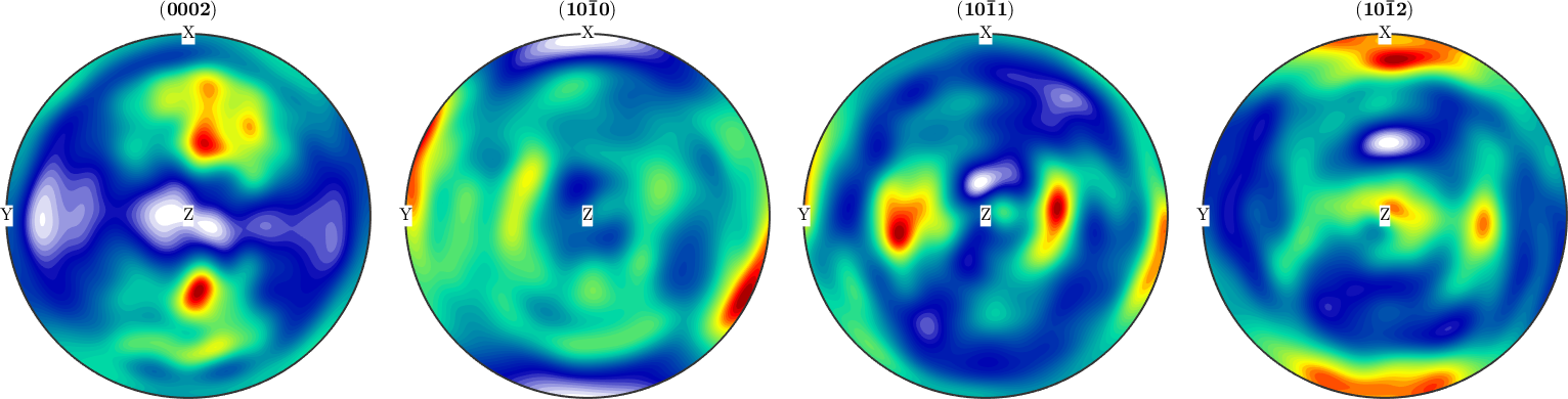
The recalculated panels are smooth fields rather than discrete dots. Compare their broad high- and low-intensity regions with the measurements above; the reconstruction should follow the structure without reproducing every point-to-point fluctuation.
calcError quantifies the same comparison. It returns one regularised relative error for each measured pole figure.
reconstructionError = calcError(pf,odf,'silent')reconstructionError =
0.0412 0.0416 0.0548 0.0418The four errors range from 0.0412 to 0.0548. They show that this ODF reproduces the measured pole figures closely after intensity scaling. A small error does not prove that the ODF is unique or physically true. ODF Reconstruction covers error measures and solver choices in detail.
Inspect the ODF
plot(odf);
mtexColorMap('LaboTeX');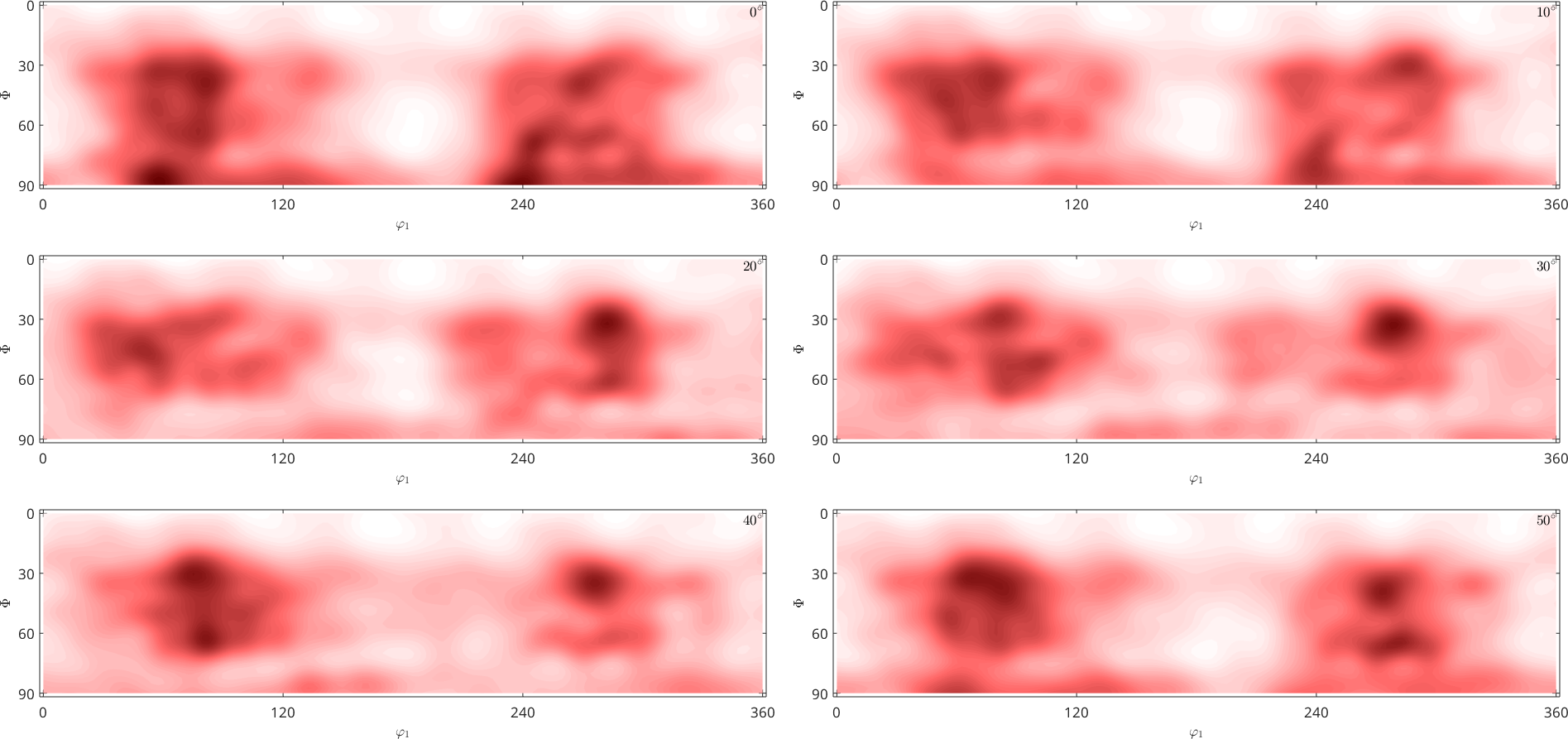
An ODF value is measured in multiples of a random distribution, mrd. A value of 1 is random density, while 10 means ten times the random density near that orientation. Compute the largest value rather than estimating it from the colour scale.
odfMaximum = max(odf,'numLocal',1)odfMaximum =
1.8528The printed maximum rounds to 1.9 mrd. The sections show broad, modest maxima rather than sharp isolated peaks, so this ZnCuTi sheet is weakly textured. Continue with ODF Analysis to choose other views, identify components, and calculate texture-dependent properties.
The maths behind the reconstruction
Let \(f(g)\) be the ODF, \(h\) a crystal-plane normal, and \(r\) a specimen direction. The corresponding pole density is the integral
\[P_h(r) = \int_{\{g:\,g h=r\}} f(g)\,\mathrm{d}g.\]
The integration set is an orientation fibre: every orientation that maps \(h\) to \(r\). This spherical Radon transform explains both why diffraction measures many crystals together and why its inversion is ambiguous.
Further reading:
- ASTM E81-96(2024), Standard Test Method for Preparing Quantitative Pole Figures, covers X-ray acquisition.
- Bunge (1969), Texture Analysis in Materials Science, develops the classical pole-figure and ODF theory.
- Matthies, Vinel and Helming (1987), Standard Distributions in Texture Analysis, gives standard distributions and the conventions used to read them.
- Hielscher and Schaeben (2008), A novel pole figure inversion method, specifies the MTEX reconstruction algorithm.
Citing this page.
This page is part of the documentation of
MTEX, a free and open
source MATLAB toolbox for analyzing and modeling crystallographic textures.
It was written by The MTEX Developers and is published at
https://mtex-toolbox.github.io/PoleFigureTutorial.html.
If you use MTEX, or reuse text or figures from this page, in your research,
please cite
F. Bachmann, R. Hielscher, H. Schaeben: Texture Analysis with MTEX - Free and Open Source Software Toolbox, Solid State Phenomena 160 (2010), 63-68. 10.4028/www.scientific.net/SSP.160.63
BibTeX
@article{bachmann2010mtex,
  author  = {F. Bachmann and R. Hielscher and H. Schaeben},
  title   = {Texture Analysis with MTEX - Free and Open Source Software Toolbox},
  journal = {Solid State Phenomena},
  volume  = {160},
  pages   = {63-68},
  year    = {2010},
  doi     = {10.4028/www.scientific.net/SSP.160.63},
  url     = {https://doi.org/10.4028/www.scientific.net/SSP.160.63}
}
Other papers describing specific MTEX methods are listed under Publications — please cite the one that best fits your application. The MTEX source code is licensed under the GNU General Public License v2.0; the text and figures of this documentation are licensed under CC BY 4.0, which permits reuse — including by automated systems — provided The MTEX Developers and this page are credited.